Should You Get a Whole-Body MRI?
I am more open to whole-body MRI than the guidelines alone might imply. It is one of the few accessible ways to collect broad structural data without radiation, and a thoughtful baseline may become more useful when compared over time. I would not treat it as the best cancer screen or let it replace targeted screening.
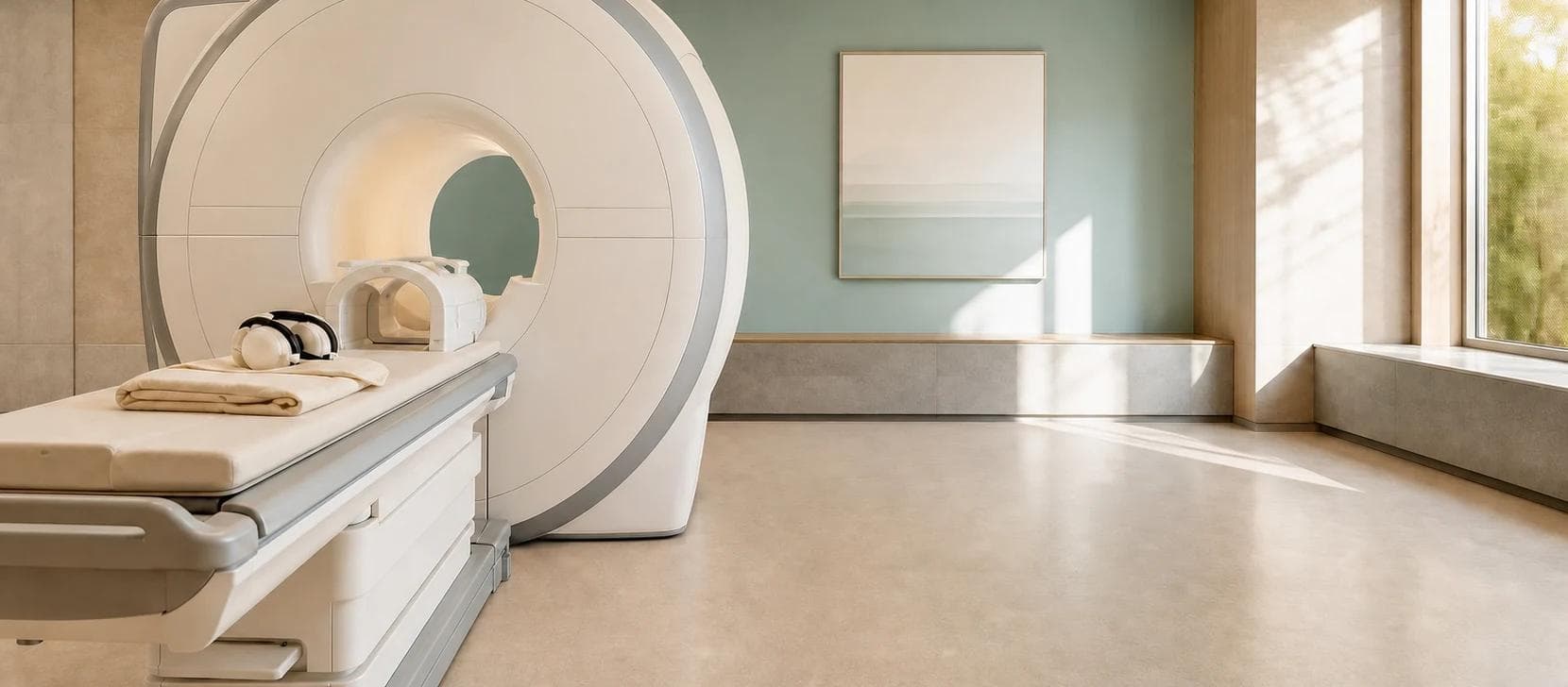
Editorial rendering, not a diagnostic study or depiction of a specific scanner model.
The short version
A structural baseline
Wearables show physiology over time. Whole-body MRI can add an episodic, radiation-free snapshot of anatomy.
Protocol decides what you see
A standard brain MRI is not the same as brain MR angiography, and vascular and organ coverage vary by program.
Not the best cancer screen
Keep mammography, colorectal screening, cervical screening, eligible-smoker lung CT, and other targeted care.
What the scan can answer
Whole-body MRI can add a structural baseline without replacing targeted screening.
Wearables show physiology over time. MRI can add an occasional anatomical snapshot, but the exact sequences decide what it can and cannot answer.
Start with the reason for scanning
Whole-body MRI is not one decision. A person with a TP53-related hereditary cancer syndrome, a person with new neurologic symptoms, and an asymptomatic adult who wants a broad baseline are asking three different clinical questions.
Symptoms belong on a diagnostic pathway. A confirmed high-risk syndrome belongs in a specialist surveillance protocol. A broad baseline is a preference-sensitive choice: it can produce useful structural data, but uncertain findings and downstream procedures are part of the tradeoff.
Screening decision
The reason for scanning determines the right pathway.
Whole-body MRI is not a single yes-or-no decision. Symptoms, hereditary risk, and a broad structural baseline start in different places.
- 01
Do you have symptoms or a new abnormal finding?
Use a targeted diagnostic pathway.
The examination, organ, and likely diagnosis should determine the protocol. A retail screening scan is not triage.
- 02
Do you have a confirmed hereditary syndrome or specialist plan?
Follow the high-risk surveillance protocol.
Whole-body MRI may be one component. It does not replace the organ-specific tests built into that plan.
- 03
Are you asymptomatic and looking for broad baseline data?
Consider it as an elective baseline.
Keep established screening current, choose the protocol deliberately, preserve the images, and plan how future comparison or follow-up would change care.
The starting point changes the default
New symptom or abnormal finding
- My default
- Use targeted diagnostic evaluation
- Why
- A focused exam and organ-specific imaging answer the actual question better than a retail screening protocol.
Confirmed hereditary cancer syndrome
- My default
- Use the specialist surveillance protocol
- Why
- Whole-body MRI may be one planned component alongside organ-specific screening and defined follow-up.
Very strong family pattern without a diagnosis
- My default
- Start with genetics and risk assessment
- Why
- The right surveillance plan depends on the syndrome and affected organs, not on a generic scan package.
Established screening is current; wants broad baseline data
- My default
- Reasonable as an elective option
- Why
- The scan may reveal useful structural findings and create a comparison point, but its outcome benefit and ideal repeat interval are unproven.
| Starting point | My default | Why |
|---|---|---|
| New symptom or abnormal finding | Use targeted diagnostic evaluation | A focused exam and organ-specific imaging answer the actual question better than a retail screening protocol. |
| Confirmed hereditary cancer syndrome | Use the specialist surveillance protocol | Whole-body MRI may be one planned component alongside organ-specific screening and defined follow-up. |
| Very strong family pattern without a diagnosis | Start with genetics and risk assessment | The right surveillance plan depends on the syndrome and affected organs, not on a generic scan package. |
| Established screening is current; wants broad baseline data | Reasonable as an elective option | The scan may reveal useful structural findings and create a comparison point, but its outcome benefit and ideal repeat interval are unproven. |
A consumer package should never be used to delay evaluation of symptoms or replace a specialist-designed high-risk plan.
What a whole-body MRI actually does
Whole-body MRI usually combines several noncontrast sequences from the head or neck through much of the torso and pelvis, sometimes extending farther. Protocols, scan time, organ coverage, image resolution, contrast use, and radiologist expertise vary. The phrase whole body is a product label, not a guarantee that every tissue received the best possible test.
MRI avoids ionizing radiation and is strong for many soft-tissue questions. Those are real advantages. But broad coverage requires tradeoffs. A fast screening protocol is not identical to a dedicated brain, breast, cardiac, liver, pelvic, spine, or musculoskeletal MRI performed for a specific indication.
I think of whole-body MRI as an extension of wearables. Wearables show physiology repeatedly: heart rate, sleep, glucose, rhythm, temperature, movement. MRI gives an occasional anatomical snapshot. Neither is a diagnosis by itself, but a well-preserved baseline can make a future change easier to recognize and interpret.
Accessible here means increasingly available without a symptom-specific diagnostic pathway, usually as a self-pay examination. It does not mean inexpensive or equitably available.
No ionizing radiationMRI uses magnetic fields and radiofrequency energy rather than X-rays.
Usually no contrastMany commercial screening protocols avoid gadolinium, but that also changes what the study can characterize.
Protocol-dependent coverageAsk what is included, excluded, and only partly assessed.
Screening is not diagnosisAn abnormal signal often needs a dedicated study before anyone knows what it means.
What a whole-body MRI may surface by region
The useful word is may. A broad noncontrast protocol can reveal structural abnormalities across several regions in one sitting, but its sensitivity is uneven. The same program may be strong for marrow and abdominal soft tissue, adequate for larger brain or pelvic findings, and weak for small lung nodules or coronary plaque.
Coverage is also not binary. Brain included does not mean head MRA. Abdomen included does not mean liver fat was quantified. Chest included does not mean the coronary arteries or small pulmonary nodules were assessed. Ask which sequences were acquired and which questions the radiologist is actually expected to answer.
What broad coverage can and cannot tell you
Brain and nervous system
- A broad protocol may surface
- Larger masses, hydrocephalus, prior major injury, and some structural abnormalities if the head is covered.
- When another test is better
- Dedicated brain MRI for a neurologic question; head MRA or CTA for aneurysms and vessels; focused pituitary, cranial-nerve, epilepsy, or demyelination protocols when relevant.
Head and neck
- A broad protocol may surface
- Thyroid nodules, enlarged lymph nodes, and larger salivary or soft-tissue masses.
- When another test is better
- Thyroid ultrasound, ENT examination, or dedicated contrast-enhanced neck imaging for characterization.
Chest and lungs
- A broad protocol may surface
- Mediastinal or chest-wall masses, pleural fluid, and some larger lung abnormalities.
- When another test is better
- Low-dose CT for validated lung-cancer screening in eligible smokers; diagnostic chest CT for small nodules and lung parenchyma. Air and motion make the lungs a relative MRI blind spot.
Heart and major vessels
- A broad protocol may surface
- Gross cardiac or aortic abnormalities when the anatomy is included, plus possible aortic enlargement depending on coverage.
- When another test is better
- Blood pressure and risk-factor assessment first; CAC or CCTA for coronary plaque questions; echocardiography or cardiac MRI for structure and function; dedicated CTA or MRA for vascular anatomy.
Abdominal organs
- A broad protocol may surface
- Liver, kidney, adrenal, pancreatic, splenic, and other solid-organ masses or cysts, plus some biliary or abdominal structural findings.
- When another test is better
- Dedicated contrast-enhanced MRI or CT, MRCP, ultrasound, endoscopy, or laboratory evaluation when the organ or lesion is the actual question.
Pelvis
- A broad protocol may surface
- Fibroids, adnexal or ovarian masses, prostate enlargement, bladder distention, and other larger pelvic abnormalities.
- When another test is better
- Pelvic ultrasound, dedicated pelvic MRI, multiparametric prostate MRI, cystoscopy, or organ-specific screening when indicated.
Skeleton, marrow, and soft tissue
- A broad protocol may surface
- Marrow-replacing lesions, larger bone or soft-tissue masses, some fractures, inflammatory changes, and common degenerative spine findings.
- When another test is better
- Targeted MRI for focal symptoms. Whole-body MRI has established roles in multiple myeloma and selected metastatic or inflammatory disease, which is different from average-risk screening.
Body composition and metabolic anatomy
- A broad protocol may surface
- Visceral and subcutaneous fat, skeletal-muscle volume, muscle fat infiltration, and sometimes liver fat when Dixon, PDFF, or validated segmentation is included.
- When another test is better
- Waist and DXA for practical serial body-composition tracking; MRI-PDFF or MR elastography for precise liver questions. A scan that can be segmented is not the same as a program that reports validated measurements.
| Region | A broad protocol may surface | When another test is better |
|---|---|---|
| Brain and nervous system | Larger masses, hydrocephalus, prior major injury, and some structural abnormalities if the head is covered. | Dedicated brain MRI for a neurologic question; head MRA or CTA for aneurysms and vessels; focused pituitary, cranial-nerve, epilepsy, or demyelination protocols when relevant. |
| Head and neck | Thyroid nodules, enlarged lymph nodes, and larger salivary or soft-tissue masses. | Thyroid ultrasound, ENT examination, or dedicated contrast-enhanced neck imaging for characterization. |
| Chest and lungs | Mediastinal or chest-wall masses, pleural fluid, and some larger lung abnormalities. | Low-dose CT for validated lung-cancer screening in eligible smokers; diagnostic chest CT for small nodules and lung parenchyma. Air and motion make the lungs a relative MRI blind spot. |
| Heart and major vessels | Gross cardiac or aortic abnormalities when the anatomy is included, plus possible aortic enlargement depending on coverage. | Blood pressure and risk-factor assessment first; CAC or CCTA for coronary plaque questions; echocardiography or cardiac MRI for structure and function; dedicated CTA or MRA for vascular anatomy. |
| Abdominal organs | Liver, kidney, adrenal, pancreatic, splenic, and other solid-organ masses or cysts, plus some biliary or abdominal structural findings. | Dedicated contrast-enhanced MRI or CT, MRCP, ultrasound, endoscopy, or laboratory evaluation when the organ or lesion is the actual question. |
| Pelvis | Fibroids, adnexal or ovarian masses, prostate enlargement, bladder distention, and other larger pelvic abnormalities. | Pelvic ultrasound, dedicated pelvic MRI, multiparametric prostate MRI, cystoscopy, or organ-specific screening when indicated. |
| Skeleton, marrow, and soft tissue | Marrow-replacing lesions, larger bone or soft-tissue masses, some fractures, inflammatory changes, and common degenerative spine findings. | Targeted MRI for focal symptoms. Whole-body MRI has established roles in multiple myeloma and selected metastatic or inflammatory disease, which is different from average-risk screening. |
| Body composition and metabolic anatomy | Visceral and subcutaneous fat, skeletal-muscle volume, muscle fat infiltration, and sometimes liver fat when Dixon, PDFF, or validated segmentation is included. | Waist and DXA for practical serial body-composition tracking; MRI-PDFF or MR elastography for precise liver questions. A scan that can be segmented is not the same as a program that reports validated measurements. |
This is a capability map, not a guarantee. Sequence selection, field of view, motion, contrast, scanner quality, and reader expertise determine what is actually reportable.
Whole-body MRI is not only a cancer-screening question
A 2026 meta-analysis of 10 studies and 9,024 asymptomatic adults found confirmed cancers in 1.57%. Most studies had moderate to serious risk of bias, protocols varied, and the studies did not establish long-term outcomes or cost-effectiveness.
The American College of Radiology says evidence is insufficient to recommend total-body MRI for people without symptoms, risk factors, or a relevant family history. It also notes there is no documented evidence that the strategy is cost-effective or prolongs life.
For average-risk adults, whole-body MRI has not been shown to reduce cancer deaths or extend life. A 1.57% detection rate is not zero, but it is not proof that routine screening improves outcomes. Whole-body MRI has detection data. Average-risk routine screening does not yet have evidence showing fewer cancer deaths, longer life, or better quality of life.
Those studies mainly ask whether broad MRI works as population cancer screening. They do not fully answer a different question: whether a high-quality structural baseline, including some noncancer and vascular findings, becomes useful when future images or symptoms need context. I think that possibility is genuinely interesting, even though its value is not yet well quantified.
Two different evidence sets
Detection is real. So is the follow-up burden.
Separate literature reviews. Not a head-to-head comparison.
Confirmed cancer detected
A detection estimate, not evidence that routine screening improves survival.
2026 meta-analysis · 10 studies · 9,024 asymptomatic adults
Critical or indeterminate incidental finding
A follow-up burden estimate, not a cancer rate or a count of dangerous disease.
2019 systematic review · 12 studies · 5,373 asymptomatic adults
Three separate standards for screening
A scan can succeed at one level without proving the next.
1. Detection
The scan marks an abnormality
Whole-body MRI clearly can do this.
2. Diagnosis
Follow-up determines what the abnormality is
Many findings remain indeterminate or turn out benign.
3. Outcome
Earlier action improves health or survival
This has not been demonstrated for routine average-risk screening.
Incidental findings are the central tradeoff
In a 2019 systematic review of 12 studies and 5,373 asymptomatic adults, the pooled prevalence of critical or indeterminate incidental findings was 32.1%. Results varied widely by protocol and study, and verification was incomplete. That number should not be read as the chance of discovering a dangerous disease. It is the chance of discovering something important-looking or unresolved enough to enter the follow-up pathway.
The cost of an incidental finding is not only money. It can mean repeat imaging, contrast, ultrasound, endoscopy, specialist visits, blood tests, biopsy, surgery, temporary restrictions, and months of uncertainty. Some of that workup is necessary. Some proves the original finding benign. The practical question is whether the expected benefit exceeds the burden for someone with your pretest risk.
Incidental does not mean irrelevant. Some unexpected findings matter. A prior baseline can also help a radiologist show that something has stayed stable, which may prevent more aggressive workup later. That value depends on keeping the source images and using a comparable protocol.
What happens after an unexpected finding
Clearly benign
- Typical next step
- Usually no action
- What remains unknown
- Whether the reporting system avoids turning trivial findings into future surveillance.
Probably benign
- Typical next step
- Repeat imaging or a dedicated study
- What remains unknown
- Whether stability will end the cascade or create serial surveillance.
Indeterminate
- Typical next step
- Targeted imaging, specialist review, or tissue sampling
- What remains unknown
- Whether the abnormality is disease, benign variation, or a technical artifact.
Concerning
- Typical next step
- Urgent diagnostic evaluation
- What remains unknown
- Whether earlier detection will materially change the treatment outcome.
| Report language | Typical next step | What remains unknown |
|---|---|---|
| Clearly benign | Usually no action | Whether the reporting system avoids turning trivial findings into future surveillance. |
| Probably benign | Repeat imaging or a dedicated study | Whether stability will end the cascade or create serial surveillance. |
| Indeterminate | Targeted imaging, specialist review, or tissue sampling | Whether the abnormality is disease, benign variation, or a technical artifact. |
| Concerning | Urgent diagnostic evaluation | Whether earlier detection will materially change the treatment outcome. |
Before scanning, decide who receives the report, who owns each recommendation, and how urgent findings are communicated.
Choose the test for the goal
Whole-body MRI is broad. Most health goals are specific. Before paying for any imaging, name the outcome you are trying to understand: coronary plaque, an occult cancer, an aneurysm, visceral fat, liver fat, bone strength, or simply a structural baseline.
The right comparison is not whole-body MRI versus doing nothing. It is whole-body MRI versus the best test for that question, plus the prevention work you could fund instead.
Choose the question first
The broadest scan is not automatically the best test.
Whole-body MRI wins on breadth. Heart, cancer, vascular, body-composition, and bone questions often need a more specific tool.
Best fit
Broad structural baseline
Best first tool
Whole-body MRI
A multi-region anatomical survey without ionizing radiation. No single targeted test offers the same breadth.
Weak fit
Heart-attack risk
Best first tool
Risk factors → CAC or CCTA when needed
A standard whole-body protocol does not measure coronary calcium, plaque burden, or stenosis.
Add-on
Cancer detection
Best first tool
Organ- and risk-specific screening
It can find some occult tumors, but mammography, cervical and colorectal screening, and eligible-smoker lung CT keep their jobs.
Protocol-dependent
Brain or aortic aneurysm
Best first tool
MRA, CTA, or risk-based ultrasound
Only count vessel screening when the written protocol includes the right angiographic sequence and coverage.
Potentially rich
Body composition
Best first tool
Waist and DXA for practical tracking
MRI can quantify regional fat and muscle when validated segmentation is actually included, not merely advertised.
Different question
Bone strength
Best first tool
DXA + fracture-risk assessment
Whole-body MRI can assess marrow and selected lesions. It does not measure validated bone-mineral density.
What to use for the question you actually have
A broad structural baseline
- Best first tools
- No single targeted test gives the same multi-region anatomical survey.
- Where whole-body MRI fits
- This is the scan's clearest elective value: broad, radiation-free structural data, provided you accept blind spots and follow-up.
Heart-attack and stroke risk
- Best first tools
- Blood pressure, ApoB or non-HDL cholesterol, glucose and diabetes risk, smoking, family history, and treatment decisions; CAC when risk is uncertain; CCTA for a coronary-anatomy question.
- Where whole-body MRI fits
- A standard whole-body MRI does not assess coronary calcium, plaque burden, or stenosis and should not be sold as a coronary-risk test.
Heart structure, valves, or function
- Best first tools
- Examination, ECG when relevant, echocardiography, rhythm monitoring, or dedicated cardiac MRI depending on the question.
- Where whole-body MRI fits
- A nongated broad scan may reveal a gross abnormality, but it is not a functional cardiac examination.
Cancer detection
- Best first tools
- Organ- and risk-specific screening: mammography, cervical HPV testing, colorectal screening, eligible-smoker low-dose CT, skin examination, and hereditary-risk assessment.
- Where whole-body MRI fits
- It can detect some otherwise unsuspected tumors and is useful in selected hereditary protocols, but it is an add-on rather than the best universal cancer screen.
Brain aneurysm or cerebral vessels
- Best first tools
- Dedicated head MRA or CTA when family history, a syndrome, symptoms, or another clinical reason makes vascular imaging appropriate.
- Where whole-body MRI fits
- Only count aneurysm screening if the written protocol includes appropriate vessel-focused sequences. Brain MRI alone is not equivalent.
Aortic aneurysm
- Best first tools
- Risk-based abdominal ultrasound; echocardiography, CTA, or MRA for thoracic or more detailed aortic assessment.
- Where whole-body MRI fits
- A broad scan may note enlargement, but coverage, cardiac motion, and measurement methods determine whether the finding is reliable.
Body composition
- Best first tools
- Waist and weight trends; DXA when lean mass, fat mass, and bone are the practical targets; dedicated MRI when regional fat or muscle detail would change a decision.
- Where whole-body MRI fits
- Potentially rich if the program quantifies visceral fat, subcutaneous fat, muscle volume, and muscle fat with validated segmentation. Many programs do not.
Fatty liver or fibrosis
- Best first tools
- Metabolic risk and liver tests, FIB-4 when appropriate, elastography for fibrosis, and MRI-PDFF or MR elastography for precise quantitative questions.
- Where whole-body MRI fits
- A broad scan may suggest steatosis, but only a suitable quantitative sequence can measure liver fat reproducibly.
Bone strength and fracture risk
- Best first tools
- DXA plus clinical fracture-risk assessment.
- Where whole-body MRI fits
- Whole-body MRI is valuable for marrow and selected bone lesions, not for validated bone-mineral-density measurement.
A focal symptom or abnormal lab
- Best first tools
- Clinical evaluation followed by the targeted test that fits the organ, anatomy, and pretest probability.
- Where whole-body MRI fits
- The retail broad-screening pathway is usually the wrong triage tool once a specific diagnostic question exists.
| Goal | Best first tools | Where whole-body MRI fits |
|---|---|---|
| A broad structural baseline | No single targeted test gives the same multi-region anatomical survey. | This is the scan's clearest elective value: broad, radiation-free structural data, provided you accept blind spots and follow-up. |
| Heart-attack and stroke risk | Blood pressure, ApoB or non-HDL cholesterol, glucose and diabetes risk, smoking, family history, and treatment decisions; CAC when risk is uncertain; CCTA for a coronary-anatomy question. | A standard whole-body MRI does not assess coronary calcium, plaque burden, or stenosis and should not be sold as a coronary-risk test. |
| Heart structure, valves, or function | Examination, ECG when relevant, echocardiography, rhythm monitoring, or dedicated cardiac MRI depending on the question. | A nongated broad scan may reveal a gross abnormality, but it is not a functional cardiac examination. |
| Cancer detection | Organ- and risk-specific screening: mammography, cervical HPV testing, colorectal screening, eligible-smoker low-dose CT, skin examination, and hereditary-risk assessment. | It can detect some otherwise unsuspected tumors and is useful in selected hereditary protocols, but it is an add-on rather than the best universal cancer screen. |
| Brain aneurysm or cerebral vessels | Dedicated head MRA or CTA when family history, a syndrome, symptoms, or another clinical reason makes vascular imaging appropriate. | Only count aneurysm screening if the written protocol includes appropriate vessel-focused sequences. Brain MRI alone is not equivalent. |
| Aortic aneurysm | Risk-based abdominal ultrasound; echocardiography, CTA, or MRA for thoracic or more detailed aortic assessment. | A broad scan may note enlargement, but coverage, cardiac motion, and measurement methods determine whether the finding is reliable. |
| Body composition | Waist and weight trends; DXA when lean mass, fat mass, and bone are the practical targets; dedicated MRI when regional fat or muscle detail would change a decision. | Potentially rich if the program quantifies visceral fat, subcutaneous fat, muscle volume, and muscle fat with validated segmentation. Many programs do not. |
| Fatty liver or fibrosis | Metabolic risk and liver tests, FIB-4 when appropriate, elastography for fibrosis, and MRI-PDFF or MR elastography for precise quantitative questions. | A broad scan may suggest steatosis, but only a suitable quantitative sequence can measure liver fat reproducibly. |
| Bone strength and fracture risk | DXA plus clinical fracture-risk assessment. | Whole-body MRI is valuable for marrow and selected bone lesions, not for validated bone-mineral-density measurement. |
| A focal symptom or abnormal lab | Clinical evaluation followed by the targeted test that fits the organ, anatomy, and pretest probability. | The retail broad-screening pathway is usually the wrong triage tool once a specific diagnostic question exists. |
A good whole-body MRI program should be willing to tell you when another test is better. Breadth is useful only when it is not confused with depth.
Body composition may be one of the more useful add-ons
MRI can separate visceral fat, subcutaneous fat, skeletal-muscle volume, muscle fat infiltration, and other tissue compartments more directly than a scale. In a 2026 study of more than 66,000 adults, age-, sex-, and height-normalized MRI measurements added prognostic information for diabetes, cardiovascular events, and mortality beyond traditional risk factors.
That makes the underlying data interesting. It does not mean every retail body-composition report is validated or that repeating the scan improves outcomes. The value depends on the actual sequence, the segmentation model, the reference population, measurement reproducibility, and whether the number changes a decision.
For routine tracking, waist, strength, performance, labs, and DXA may be easier to repeat and act on. MRI becomes more compelling when regional fat distribution, muscle volume, muscle quality, or liver fat is the question and the program provides reproducible quantitative outputs.
Measurement needs a method
“Body composition included” is not enough.
| Measure | Broad MRI | What makes it quantitative | Practical comparator |
|---|---|---|---|
| Visceral and subcutaneous fat | Potentially quantifiable | Validated whole-volume segmentation and a repeatable acquisition | Waist for simple serial tracking; DXA for total and regional fat |
| Muscle amount and quality | Area, volume, and fat infiltration may be measured | Defined landmarks or whole-volume segmentation plus validated water-fat separation | DXA lean mass; strength and performance answer function |
| Liver fat | Not from generic images alone | MRI-PDFF or another validated quantitative fat sequence | Metabolic risk and labs first; MRI-PDFF when precision changes care |
| Bone density | Not measured | MRI may show marrow or lesions, not bone-mineral density | DXA plus clinical fracture-risk assessment |
Visceral and subcutaneous fat
- Broad MRI
- Potentially quantifiable
- What makes it quantitative
- Validated whole-volume segmentation and a repeatable acquisition
- Practical comparator
- Waist for simple serial tracking; DXA for total and regional fat
Muscle amount and quality
- Broad MRI
- Area, volume, and fat infiltration may be measured
- What makes it quantitative
- Defined landmarks or whole-volume segmentation plus validated water-fat separation
- Practical comparator
- DXA lean mass; strength and performance answer function
Liver fat
- Broad MRI
- Not from generic images alone
- What makes it quantitative
- MRI-PDFF or another validated quantitative fat sequence
- Practical comparator
- Metabolic risk and labs first; MRI-PDFF when precision changes care
Bone density
- Broad MRI
- Not measured
- What makes it quantitative
- MRI may show marrow or lesions, not bone-mineral density
- Practical comparator
- DXA plus clinical fracture-risk assessment
What the scan should never replace
The first opportunity cost is usually not another scan. It is the proven or guideline-supported prevention that never happened because the broad MRI felt more comprehensive.
Blood pressure, lipid and ApoB assessment when useful, glucose, smoking, alcohol, sleep, exercise, and medication review.
Breast, cervical, colorectal, and lung-cancer screening when eligible.
Vaccination, skin evaluation based on risk, and bone-density testing when indicated.
Genetic counseling when family history suggests a hereditary cancer syndrome.
Targeted diagnostic workup for symptoms, examination findings, or abnormal laboratory results.
When whole-body MRI can be reasonable
The strongest evidence is still planned surveillance in selected high-risk populations where multiple tumor types matter and avoiding repeated radiation is valuable. Li-Fraumeni syndrome and some other cancer-predisposition syndromes are the clearest examples, although the exact protocol should come from a genetics or oncology team.
That is not the only reasonable use. I also think a one-time baseline can make sense for a high-agency adult who wants broad structural data, understands the uncertainty, has established screening current, can afford the downstream work, and has a clinician prepared to manage every result.
A confirmed hereditary syndrome with an established surveillance protocol.
A specialist-defined high-risk pathway involving multiple organ systems.
A specific unresolved question that targeted evaluation has not answered and for which the result changes care.
A deliberate structural baseline that can be preserved and compared if symptoms, risk, or future findings change.
A preference-sensitive elective decision made after established screening is current and the follow-up burden is acceptable.
When I would usually skip it
I would usually skip elective whole-body MRI when the main goal is reassurance, when health anxiety is likely to worsen with uncertain findings, when guideline-supported screening is overdue, or when there is no clear clinician to own the report. I would also skip it when paying for the scan would crowd out medications, dental care, sleep-apnea testing, physical therapy, vaccination, or ordinary primary and preventive care.
A retail scan is especially poor triage for active symptoms. A symptom changes the pretest probability and the appropriate protocol. If you have a new lump, focal weakness, unexplained bleeding, chest symptoms, persistent fever, major unexplained weight loss, or another red flag, seek clinical evaluation rather than buying a broad screen.
The result would not change anything you are willing to do.
No one has agreed to review the report and coordinate follow-up.
You are likely to pursue every tiny finding regardless of risk.
You may ignore a negative study despite symptoms or overdue screening.
The cost displaces care with clearer expected benefit.
If you choose the scan, arrange follow-up first
Do not schedule the scan before you understand the handoff. Before you pay, confirm what was imaged, who read it, how uncertainty is graded, how urgent results reach you, and who will manage every follow-up recommendation.
Name the clinician who will review the full report, not only the summary call.
Ask how same-day and urgent findings are communicated and documented.
Ask which organs are excluded or only partly assessed, including breast, lungs, heart, bowel, and skin.
Ask what proportion of clients receive a recommendation for more imaging or a specialist visit.
Ask whether dedicated follow-up imaging and specialist care are included, discounted, or entirely separate.
Keep the original images and radiology report in a form your clinicians can access.
Decide in advance how you will respond to a probably benign or indeterminate finding.
How to compare whole-body MRI programs
Questions worth asking before you pay
What exact anatomy is covered?
- Why it matters
- Whole body is not a standardized promise. Head, breasts, heart, lungs, extremities, and bowel coverage vary.
Does brain coverage include MR angiography?
- Why it matters
- Routine brain sequences and vessel-focused MRA answer different questions. If aneurysm detection matters to you, get the exact protocol in writing.
How is the aorta assessed?
- Why it matters
- Ask which segments are covered, how diameter is measured, and whether a dedicated vascular study would still be needed for your risk.
What scanner strength and sequences are used?
- Why it matters
- Field strength alone does not prove quality; the protocol and reader expertise matter.
Is contrast used?
- Why it matters
- Contrast can improve selected diagnostic questions but introduces a different risk and consent discussion.
Who reads the study?
- Why it matters
- Ask about radiologist training, subspecialty review, double reading, and quality assurance.
How are findings categorized?
- Why it matters
- A useful report distinguishes clearly benign, probably benign, indeterminate, and concerning findings.
What is the follow-up rate?
- Why it matters
- This is a better preview of downstream burden than the company's number of conditions screened.
How are urgent results handled?
- Why it matters
- A marketing consultation is not the same as a clinical escalation pathway.
Can my physician receive images and the full report?
- Why it matters
- Portability prevents the scan from becoming an isolated data silo.
Can a future scan be compared reliably?
- Why it matters
- Longitudinal value is stronger when the images, sequences, positioning, and measurements are comparable.
What happens if I cannot tolerate the scan?
- Why it matters
- Claustrophobia, noise, pain, and scan duration can limit image quality or completion.
How are implants and pregnancy screened?
- Why it matters
- MRI safety depends on the device, material, sequence, body region, and clinical indication.
What is the total out-of-pocket cost?
- Why it matters
- The scan, blood tests, contrast, consultation, follow-up imaging, and specialist care may be billed separately.
What evidence supports each add-on?
- Why it matters
- Body composition, brain-age scores, AI outputs, and cardiac or metabolic extras should be judged separately.
| Question | Why it matters |
|---|---|
| What exact anatomy is covered? | Whole body is not a standardized promise. Head, breasts, heart, lungs, extremities, and bowel coverage vary. |
| Does brain coverage include MR angiography? | Routine brain sequences and vessel-focused MRA answer different questions. If aneurysm detection matters to you, get the exact protocol in writing. |
| How is the aorta assessed? | Ask which segments are covered, how diameter is measured, and whether a dedicated vascular study would still be needed for your risk. |
| What scanner strength and sequences are used? | Field strength alone does not prove quality; the protocol and reader expertise matter. |
| Is contrast used? | Contrast can improve selected diagnostic questions but introduces a different risk and consent discussion. |
| Who reads the study? | Ask about radiologist training, subspecialty review, double reading, and quality assurance. |
| How are findings categorized? | A useful report distinguishes clearly benign, probably benign, indeterminate, and concerning findings. |
| What is the follow-up rate? | This is a better preview of downstream burden than the company's number of conditions screened. |
| How are urgent results handled? | A marketing consultation is not the same as a clinical escalation pathway. |
| Can my physician receive images and the full report? | Portability prevents the scan from becoming an isolated data silo. |
| Can a future scan be compared reliably? | Longitudinal value is stronger when the images, sequences, positioning, and measurements are comparable. |
| What happens if I cannot tolerate the scan? | Claustrophobia, noise, pain, and scan duration can limit image quality or completion. |
| How are implants and pregnancy screened? | MRI safety depends on the device, material, sequence, body region, and clinical indication. |
| What is the total out-of-pocket cost? | The scan, blood tests, contrast, consultation, follow-up imaging, and specialist care may be billed separately. |
| What evidence supports each add-on? | Body composition, brain-age scores, AI outputs, and cardiac or metabolic extras should be judged separately. |
Program pricing and bundled features change. Compare the current written protocol and follow-up terms, not only an advertised package name.
AI and add-ons do not solve the outcome question
AI can help with image acquisition, segmentation, quality control, measurement, and prioritization. It can also generate more flagged regions, proprietary scores, and apparent precision. More output is useful only when the output is validated, clinically interpretable, and linked to a better decision.
Longitudinal measurement is one place AI may become genuinely useful: segmenting the same structures and showing credible change rather than asking a person to compare two reports by eye. But every add-on remains a separate product claim. Ask what was validated, how often it is wrong, whether a clinician can inspect the measurement, and what action changes.
A baseline becomes useful when the comparison is real
A prior image can answer a question that a single report cannot: was this structure already present, and has it changed? That is the strongest first-principles case for keeping the original images rather than treating the scan as a one-time reassurance product.
The comparison still has to be technically credible. Scanner, sequence, slice thickness, positioning, breath-hold, segmentation model, and reader measurement can all create apparent change. A finding that is two millimeters larger is not automatically biologic growth.
From scan to signal
A baseline is not the answer. It is the first frame.
00 · CAPTURE
Keep the real study
Preserve DICOM images, the full report, exact sequences, and any quantitative measurements. A summary PDF is not a reusable baseline.
01 · REPEAT
Make it comparable
Match anatomy, sequence, positioning, and measurement method. Otherwise scanner and protocol variation can masquerade as biologic change.
02 · INTERPRET
Separate change from noise
Stable
Often the most reassuring use of a prior image.
Meaningful change
New, clearly larger, or clinically different enough to alter care.
Not comparable
A technical difference, uncertain measurement, or different field of view.
Preserve the DICOM images and full report, not only a consumer summary.
Use comparable anatomy, sequences, and measurement methods on repeat imaging.
Separate stable findings, clearly new findings, meaningful growth, and measurement noise.
Let a defined clinical question or surveillance protocol set the interval; average-risk annual scanning is not established.
Make sure a clinician can review both studies side by side and decide whether the change is actionable.
Where this fits in prevention care
I am more open to whole-body MRI than population-screening guidelines alone might suggest. It is one of the few accessible, radiation-free ways to collect broad data about what is happening inside the body. I think of it as an extension of wearables: not continuous physiology, but an occasional structural baseline that may become more informative when it changes.
I would not call it the best cancer screen. I would keep every targeted test that does its job better. But after those are current, I think it can be a reasonable way to explore anatomy, capture a baseline, and learn over time which findings matter and which do not.
Keep symptom evaluation, family-history work, hereditary-risk assessment, and established screening separate.
Choose the protocol for what you actually want to know, including brain and vascular coverage.
Preserve the original images so future comparison is possible.
Repeat only when the comparison has a purpose, not because an annual subscription says it is time.
How I’d decide
Who it’s for
Adults who want a broad structural baseline, understand the uncertainty, have established screening current, can manage follow-up, and have a clinician who can review the full report. Hereditary risk and specialist surveillance remain stronger evidence-based indications.
Who should skip it
Skip the retail screening pathway if you have new symptoms, a new lump, neurologic change, unexplained bleeding, major weight loss, persistent fever, or another clinical red flag; those need targeted evaluation. I would also skip it if uncertain findings are likely to worsen health anxiety or no one will own follow-up.
Measure before / after
Before scanning, define the purpose, exact anatomy and sequences, whether brain MR angiography or aortic assessment is included, what the protocol excludes, who reads the study, how images will be preserved for comparison, and who owns follow-up.
What I’d do first
After guideline-supported screening and prevention are current, I would be open to one high-quality whole-body MRI as a broad structural baseline. I would choose the protocol deliberately, preserve the source images, and repeat it only when a comparable scan could answer a real question about change over time.
What would change my mind
A strong program with transparent coverage, high-quality radiology, portable images, consistent repeat protocols, and clinician-owned follow-up makes a baseline more useful. Better prospective evidence on which findings predict meaningful disease, which changes matter, and what repeat interval improves decisions would make longitudinal use much stronger.
Frequently Asked Questions
Can whole-body MRI detect cancer early?
It can detect some cancers and important abnormalities before symptoms appear, but performance varies by organ, tumor type, size, protocol, and reader. Early detection is not automatically beneficial; the unanswered average-risk question is whether routine screening reduces cancer deaths or improves quality of life enough to outweigh false positives and downstream procedures.
Does whole-body MRI use radiation?
No ionizing radiation is used. MRI uses magnetic fields and radiofrequency energy. That is an advantage, especially in surveillance programs that require repeated imaging, but it does not eliminate incidental findings, missed disease, safety screening, or follow-up burden.
Does a negative scan mean I am healthy?
No. It means the protocol did not identify a reported concerning finding at that time. Microscopic disease, fast-growing disease, organ-specific blind spots, and conditions better detected by examination, laboratory testing, endoscopy, mammography, CT, or another dedicated study can still be present.
Can whole-body MRI detect brain or aortic aneurysms?
Sometimes, but the protocol decides what can be seen. Brain aneurysm assessment generally uses dedicated head MR angiography or CT angiography; a routine brain MRI is not the same study as angiography. A broad scan may show aortic enlargement, but validated abdominal-aortic screening uses ultrasound in eligible adults, and higher-risk vascular questions may need dedicated CTA or MRA.
Is whole-body MRI a good heart-health screen?
Not for the most common prevention question. A standard whole-body MRI does not measure coronary calcium, plaque burden, stenosis, rhythm, valve function, or exercise capacity. Heart-health decisions usually start with blood pressure, ApoB or other lipids, glucose risk, smoking, family history, and treatment; CAC, CCTA, echocardiography, rhythm monitoring, or cardiac MRI answer specific imaging questions.
Is whole-body MRI useful for body composition?
Potentially. Suitable MRI sequences and validated segmentation can quantify visceral fat, subcutaneous fat, muscle volume, muscle fat infiltration, and sometimes liver fat. The important distinction is whether a program actually reports reproducible measurements. For ordinary tracking, waist, performance, and DXA may be easier to repeat and act on.
Should I get a whole-body MRI every year?
There is no evidence-based default annual interval for average-risk adults. Longitudinal comparison may become valuable, but only if the protocols are comparable and the change would affect a decision. Repeat timing should follow a high-risk surveillance plan, a specific finding, or a defined question, not a subscription cadence.
What is the risk of false positives?
The exact rate varies by protocol and by how studies define and verify findings. A 2019 systematic review found a pooled 32.1% prevalence of critical or indeterminate incidental findings in asymptomatic adults, with substantial heterogeneity and incomplete verification. That is not a cancer rate; it is a warning that follow-up is common enough to plan for.
Is whole-body MRI safe during pregnancy?
Pregnancy does not make all MRI unsafe, but elective whole-body screening is not the right framework. Imaging during pregnancy should answer a specific clinical question, use an appropriate protocol, and involve the obstetric and radiology teams. Gadolinium generally requires a separate, indication-specific risk-benefit decision.
What if I have metal or an implanted device?
Tell the imaging center before scheduling. Safety depends on the exact device or material, manufacturer labeling, implantation details, field strength, body region, and protocol. Never assume that a device is safe because a prior scan was uneventful.
Is whole-body MRI covered by insurance?
Elective screening without a medical indication is usually self-pay in the United States. Coverage for follow-up imaging, specialists, or procedures depends on the finding, insurance plan, and medical-necessity rules. Ask about the entire likely cascade, not only the scan price.
Is AI-read whole-body MRI better?
Not automatically. AI may improve acquisition, measurement, workflow, or detection for a validated task. It can also increase flagged findings or generate scores with unclear clinical meaning. Ask for validation, error rates, clinician oversight, and the action tied to each output.
Would you get one yourself?
Yes, I am open to a high-quality one-time scan as a broad structural baseline after my targeted screening is current. I would want the exact brain and vascular coverage, the source images, and a clinician who can manage the report. I might repeat it if a comparable scan could answer a real question about change, but not automatically every year.
References & citations
- 1.American College of Radiology. ACR Statement on Screening Total Body MRI, 2023.
- 2.Martins da Fonseca et al. Whole-Body MRI for Opportunistic Cancer Detection in Asymptomatic Individuals: A Systematic Review and Meta-Analysis. European Radiology, 2026. PMID 40884613.
- 3.Kwee and Kwee. Whole-Body MRI for Preventive Health Screening: A Systematic Review of the Literature. Journal of Magnetic Resonance Imaging, 2019. PMID 30932247.
- 4.Dacoregio et al. Baseline Surveillance in Li-Fraumeni Syndrome Using Whole-Body MRI: A Systematic Review and Updated Meta-Analysis. European Radiology, 2025. PMID 39075300.
- 5.Greer et al. Clinical Applications and Controversies of Whole-Body MRI. American Journal of Roentgenology, 2023. DOI 10.2214/AJR.22.28229.
- 6.Schneider et al. Li-Fraumeni Syndrome. GeneReviews, updated regularly. NCBI Bookshelf.
- 7.U.S. Preventive Services Task Force. A and B Recommendations: evidence-based preventive services by age and risk.
- 8.American College of Radiology. MR Safety Resources and Manual on MR Safety, 2026.
- 9.Radiological Society of North America. Whole-Body MRI Expands, Leaving Patients Weighing Risks, July 2026.
- 10.American College of Radiology and Radiological Society of North America. Cerebrovascular Diseases: Aneurysm, Vascular Malformation, and Subarachnoid Hemorrhage.
- 11.U.S. Preventive Services Task Force. Abdominal Aortic Aneurysm: Screening recommendation.
- 12.Padhani et al. Present and Future of Whole-Body MRI in Metastatic Disease and Myeloma. European Radiology Experimental, 2024.
- 13.Langner et al. Body Composition in the General Population: Whole-Body MRI-Derived Reference Curves From Over 66,000 Individuals. Radiology, 2026. PMID 42084507.
- 14.RSNA Quantitative Imaging Biomarkers Alliance. MRI-Based Proton Density Fat Fraction of the Liver Profile, consensus-maintenance version, 2024.
- 15.American Heart Association. Coronary Artery Calcium Test, reviewed 2026.
- 16.American College of Radiology and Radiological Society of North America. Bone Density Scan (DXA or DEXA).
Related Guides
Before you book the scan
Choose the protocol before you choose the scanner.
Broad data becomes more useful when the coverage is clear, future scans are comparable, and every unexpected finding has a clinical handoff.